2019-45 / DECEMBER 30 (CONTRIBUTOR: JOANNA ROGALA )
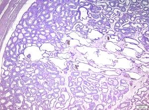
A 46 y/o male with a testicular mass undergoes orchiectomy. On gross examination a well demarcated 1,5 cm tumor with spongy appearance confined to testis is seen.
Previous
Next
Quiz
1. What is the correct diagnosis?
a. Cystic transformation of rete testis
b. Yolk sac tumor (Macrocystic or polyvesicular vitelline pattern)
Cystic transformation of rete testis (CTRT) is a benign lesion occurring in an adult man, often caused by obstruction of epididymis or spermatic cord by one of following:
Other associations include malformations (cryptorchism), hormonal alterations (liver cirrhosis) and hemodialysis due to chronic renal insufficiency.
Histologic picture include conglomerate of multiple, thin walled cysts, lined by flat, clearly benign looking rete testis type epithelium. Positive for EMA, PAX-8; negative for germ cell markers (SALL4, OCT3/4, AFP).
Yolk Sac Tumour – malignant neoplasm, with tumour cells showing mild to moderate atypia and frequent mitotic figures. In adult population extremely rare occurs in a pure form and if, there is rather mixture of patterns than one, single pattern. Associated with germ cell neoplasia in situ. Prognosis is rather poor, correlated with clinical stage and presence of lymphovascular invasion. In contrast, in children it’s the most common pure germ cell tumour; macrocystic pattern being one of the most common ones. In adjacent testicular tissue there is no germ cell neoplasia in situ. Prognosis is excellent. Positive for cytokeratin, AFP; negative for CD30, D2-40, OCT3/4, calretinin
Cystic dysplasia of the rete testis is a benign congenital lesion diagnosed in children, frequently associated with ipsilateral renal agenesis and dysplasia. Histologic picture is similar to that of CTRT. Positive for EMA, PAX-8; negative for germ cell markers (SALL4, AFP).
Adenomatoid tumor is a benign paratesticular neoplasm of mesothelial origin, occurring most commonly in epididymis. Rarely, intratesticular location is possible. Microscopic picture shows well demarcated tumour composed of irregular tubules, nests, microcysts with flat to cuboidal epithelioid cells. So-called bridging strands are always present, formed by flat cells of mesothelial origin. Cytoplasmic vacuoles may be present. Positive for cytokeratin, calretinin.
Nistal M, Mate A, Paniagua R. Cystic transformation of the rete testis. Am J Surg Pathol. 1996 Oct; 20(10):1231-9.
Jeyaratnam R, Bakalinova D.Cystic dysplasia of the rete testis: a case of spontaneous regression and review of published reports. Urology. 2010 Mar;75(3):687-90
Pascual Mateo C, Fernández González I, Luján Galán M, Rodríguez García N, Espinales Castro G, Berenguer Sánchez A. Cystic ectasia of the rete testis. Arch Esp Urol. 2006 Jan-Feb;59(1):55-8.
NSure ogales FF Jr.,Matilla A, Nogales-Ortiz F, L.Galera-Davidson H.Yolk sac tumors with pure and mixed polyvesicular vitelline patterns. Human Pathology, Volume 9, Issue 5, September 1978, Pages 553-566.