CASE OF THE WEEK
2019-37 / NOVEMBER 4
(CONTRIBUTOR: NERIMEN GOKDEN)
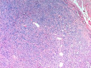
70 year old male with a bladder mass. CT with contrast showed a 2 cm left sided well-circumscribed bladder mass in the wall. Robotic partial cystectomy was performed. Pictures: 1-3 H&E’s, 4- CD34, 5- STAT6
Quiz
What is the correct diagnosis?
a. Neurofibroma
b. Hemangioma/vascular malformation
c. PEComa
d. Solitary fibrous tumor
1. d
1. Solitary fibrous tumor
This case shows intramural nodular mass in the bladder with microscopic features of angulated (hemangiopericytic) blood vessels, variable cellularity, less organized fascicles, and collagenized areas. Nuclear pleomorphism and mitotic activity were not found. Tumor cells expressed cytoplasmic CD34, and nuclear STAT6 which is a surrogate marker for the fusion involving NAB2 and STAT6, and is characteristic of Solitary Fibrous Tumor (SFT).
Neurofibromas demonstrates spindle cells with small tapered or wavy nuclei, randomly distributed bundles of collagen, and S100 positivity. In this case S100 was negative. Hemangiomas are designated as vascular malformation under recent classification and composed of blood vessels lined by cytologically bland endothelial cells. They commonly express CD34 and CD31. PEComas display variable histologic features, composed of spindled cells, epithelioid cells, and nested growth pattern. Focally tumor cells present within the walls of blood vessels. Typically tumor cells coexpress smooth muscle antigen and melanocytic markers (HMB45, Melan-A and MiTF). In this case CD31, SMA, and melanocytic markers were negative.
Most of the SFTs behave in a benign fashion. Malignant potential is predicted when the tumor is large size, and nuclear pleomorphism and mitotic activity are present.
Tanaka EY, Buonfiglio VB, Manzano JP, Filippi RZ, Sadi MV. Two cases of Solitary Fibrous Tumor involving Urinary Bladder and a review of the Literature.
Case reports in Urology. 2016; 2016: Article ID 5145789.
Neriman Gokden
University of Arkansas for Medical Sciences, Little Rock, AR
gokdenneriman@uams.edu
Bladder
Bladder, solitary fibrous tumor, immunohistochemistry


